Kategorier
SIC synchrotrons

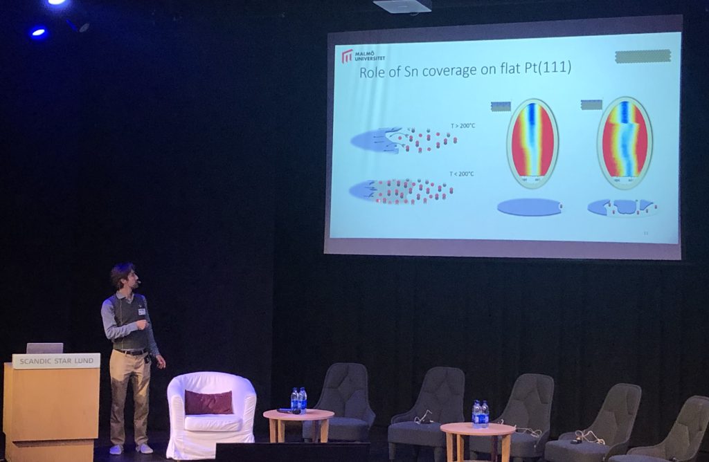
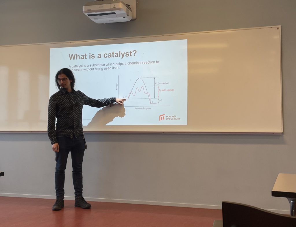
Miguel presents his thesis project in a startup seminar

Miguel Kofoed, who started his PhD in Applied Physics in November, presented his plans for his thesis work in a seminar today. Miguel is part of the PRISMAS Graduate School, a national network coordinated by the MAX IV Laboratory and funded by the EU through an MSCA Cofund project. His project is a collaboration between MaU and MAX IV, where he will work on the integration of diffraction and scattering methods at the Balder beamline, with special focus on the diffraction anomalous fine structure (DAFS) technique. DAFS can give atomic-scale information about materials like catalysts and batteries during operation, and so should help us to improve these technologies. Welcome Miguel and best of luck!
Kategorier
imaging synchrotrons

Experiments with metal foams at ESRF

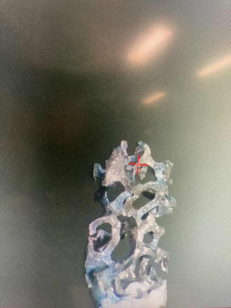
Between 14th – 18th September, Indrajeet, Pär and Johan together with researchers from Lund and Ulm Universities went to Grenoble to conduct measurements at the European Synchrotron Radiation Facility (ESRF). The purpose of the visit was to investigate the microstructural evolution of metallic foams during heat treatment using an in-situ x-ray setup. This is part of Indrajeet’s PhD project, which is part of a joint Swedish-German project funded by the Swedish Research Council. The in-situ experiments were challenging, but the results look promising. Thus, we are confident that you will hear about the results at a conference in the near future.

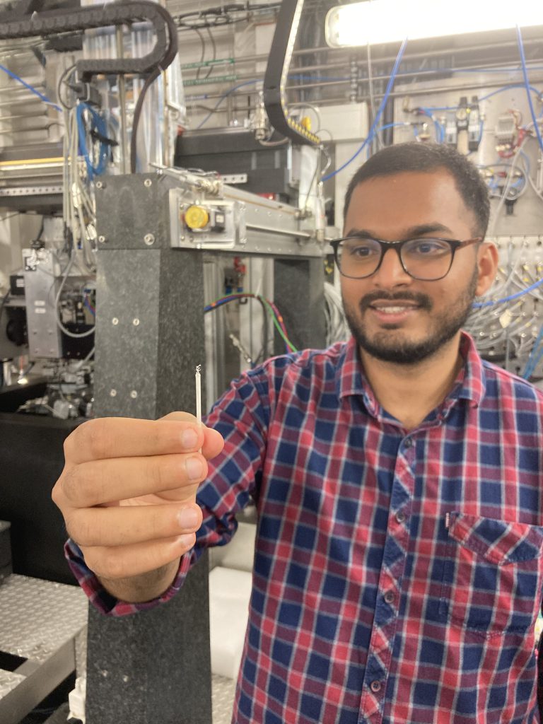
Indrajeet holding the metal foam sample before loading it into the X-ray imaging instrument.
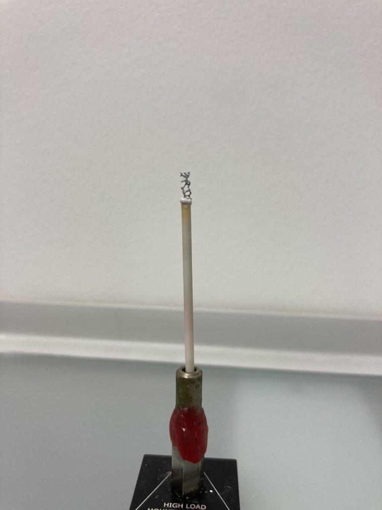
Close-up view of the sample, which is a small piece cut out from a block of metal foam.
View of the metal foam sample though the camera used for positioning in the X-ray instrument.
Kategorier
SIC synchrotrons

Electrocatalysis experiments at PETRA III

Dorotea, Harald and Lindsay were at the PETRA III synchrotron in Hamburg last week, making high-energy surface X-ray diffraction measurements on model electrocatalysts – simplified materials that should help us understand how to improve the performance of electrolyzers and fuel cells. The experiments were part of a collaboration with Prof. Gary Harlow from the Oregon Center for Electrochemistry at the University of Oregon (go Huskies!). The X-ray measurements were made at the Swedish Materials Science beamline P21. Also joining the experiment was Andrea Grespi, PhD student from Lund University.

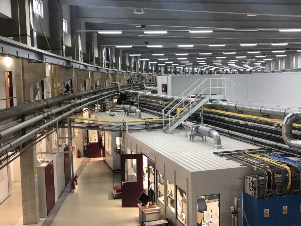
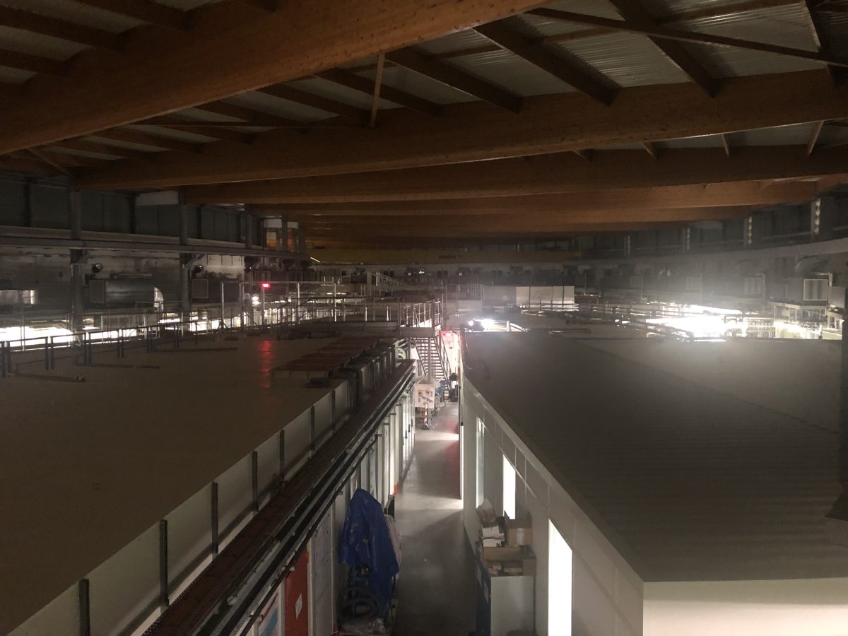
View across the “Max von Laue” ring hall at the PETRA III synchrotron, Hamburg, Germany.
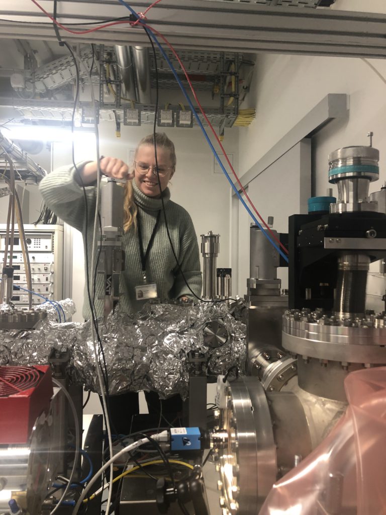
Samples were initially prepared at the DESY Nanolab (thanks Heshmat and colleagues!) before transferring to the synchrotron for measurements. Dorotea was in charge of this part of the experiment. Here she is moving the freshly-prepared sample (which you can’t see) through the ultra-high vacuum system.
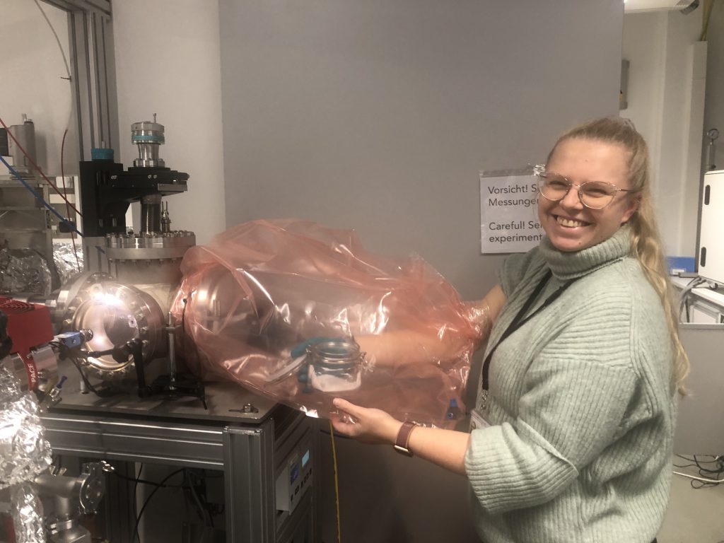
The samples are very delicate, so we try to avoid exposure to too much air. Here’s the improvised solution for this experiment: a bag sealed around the entry door to the vacuum system. The bag is filled with dry nitrogen and the sample transferred into an airtight jar, which we can then take to a proper glove box to load into the electrochemistry cell.
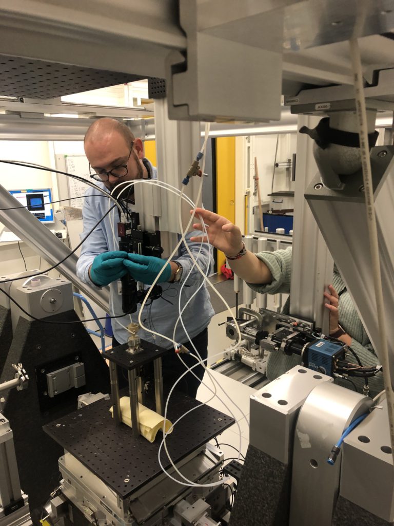
After getting the sample loaded into the electrochemistry cell (the beige thing on the pedestal here with tubes coming out), this is mounted at the X-ray beamline for measurements. Here you see the setup almost ready; Gary is adjusting the camera we use to monitor the surface of the sample during measurements.
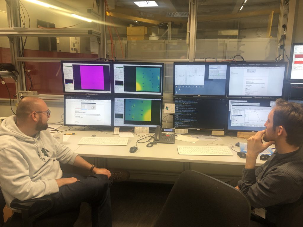
Gary and Harald watching as the X-ray diffraction data come in.
X-ray diffraction pattern from the sample, which is a material only a few atomic layers thick. The pattern of spots tells us about the atomic structure, and we can follow changes in real time while we change the conditions.
Kategorier
imaging synchrotrons

Surface diffraction imaging experiments at DanMAX

Johan and Indrajeet were at the MAX IV beamline DanMAX last week for an experiment using surface X-ray diffraction to map grains on polycrystalline surfaces. The new technique they are helping to develop is called “surface X-ray diffraction tomography”, and will enable surface structural characterization on complex samples under in situ conditions. The project is led by Johan Gustafson at Lund University.

Kategorier
SIC synchrotrons

Surface X-ray diffraction experiments at SOLEIL

Lindsay and Harald were at the SOLEIL synchrotron last week to perform measurements at the beamline SixS. They used X-rays to study the surface structure of an alloy of platinum with tin. The experiment was part of a collaboration with Lund University and the University of Bremen.

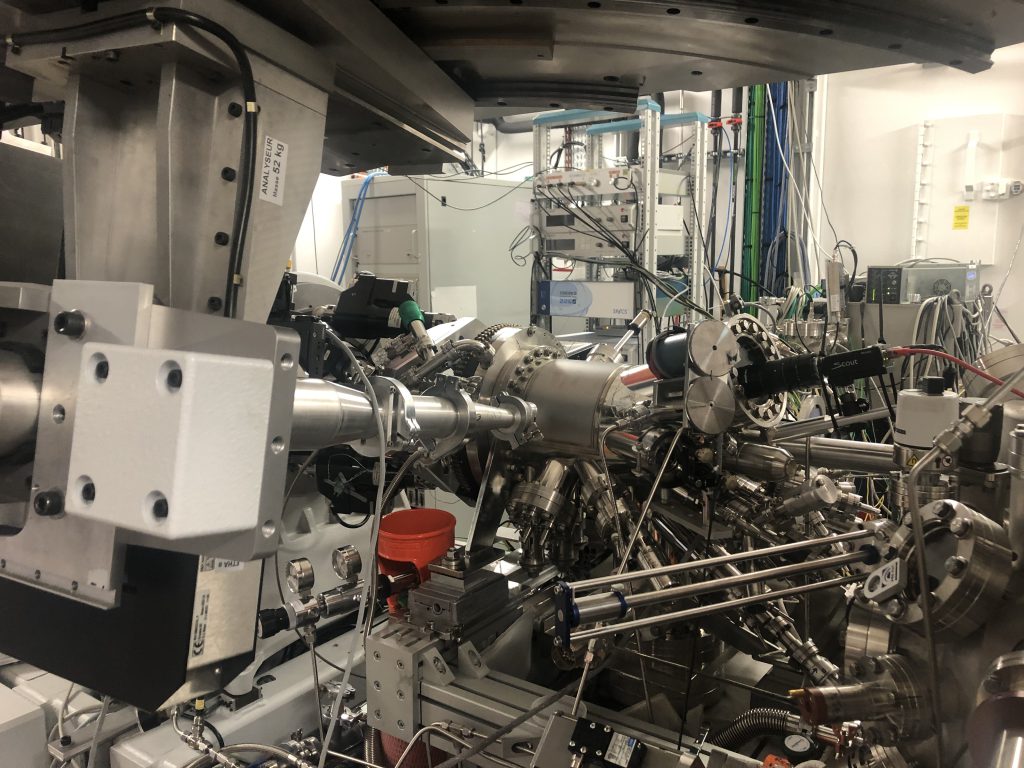
The X-ray diffraction instrument at SixS used to study surfaces in ultra-high vacuum.
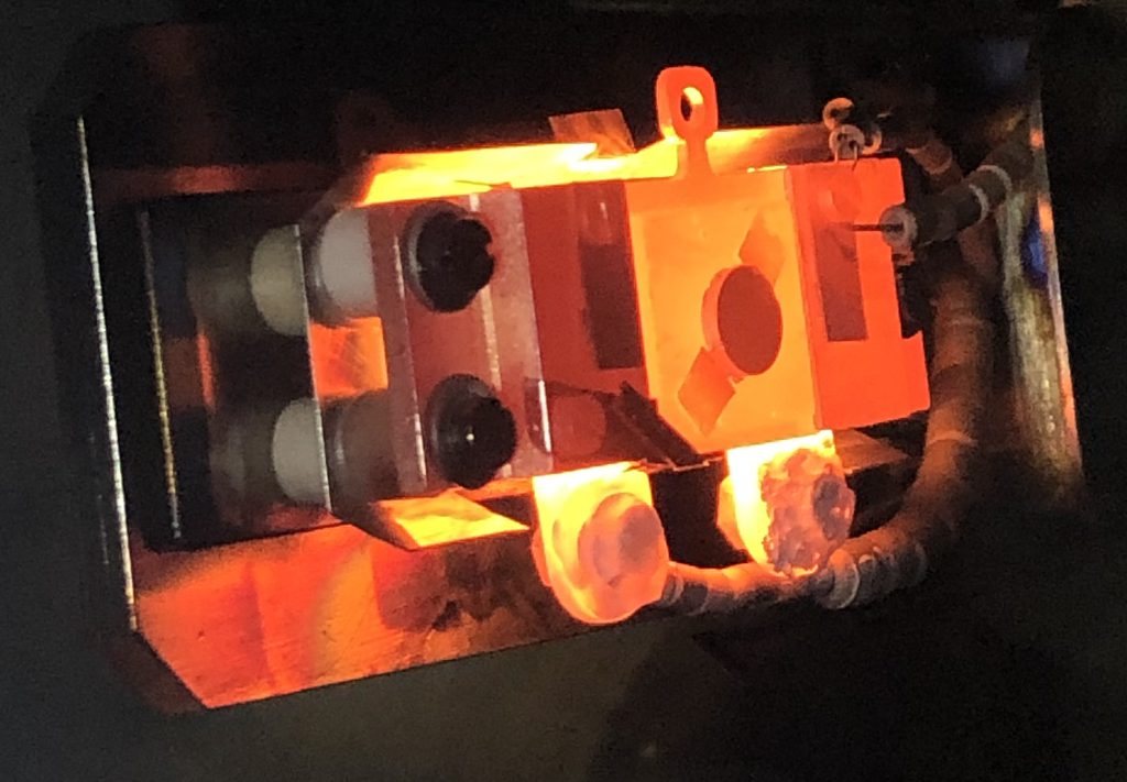
The sample being heat-treated in the vacuum chamber. It’s the red-orange disc in the middle of the rectangular yellow-orange plate.
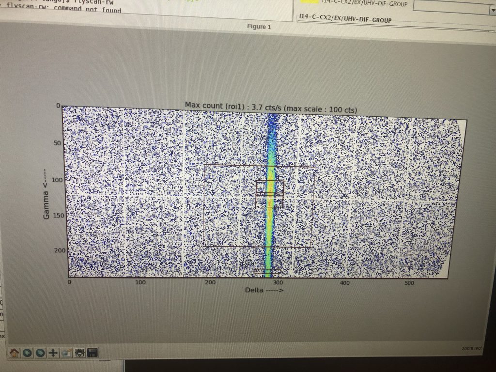
Surface X-ray diffraction signal. The vertical streak is called a ‘rod’, and is characteristic for X-ray scattering from two-dimensional structures.
Kategorier
imaging synchrotrons

Bragg CDI experiments at NanoMAX

Martin, Johan, Pär, and Mattias were at MAX IV last week performing measurements at the NanoMAX beamline. The goal of the experiment was to test the application of Bragg coherent diffraction imaging (CDI) to study precipitates in metal alloys.

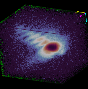
3D map of a coherent Bragg reflection from a precipitate in an aluminum alloy
Kategorier
SIC synchrotrons

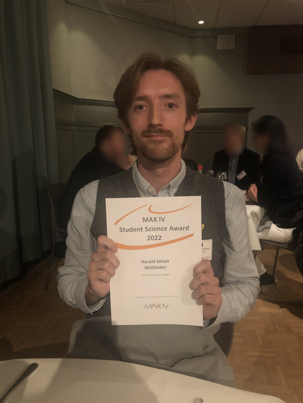
MAX IV Student Science Award to Harald

Harald Wallander won the Student Science Award at the annual MAX IV User’s Meeting. He presented his work looking at platinum-tin surface alloys with ambient-pressure XPS, based on experiments done at the HIPPIE beamline. Congrats Harald!

Harald presenting his work at the plenary session.
This is the MaU logo